We present a 23-year-old female patient diagnosed with Shone syndrome (SS), consisting of a bicuspid aortic valve, severe coarctation of the aorta (CoA), hypoplastic aortic arch, and parachute mitral valve (PMV). One week after birth, she underwent surgical repair of CoA using a side-to-side anastomosis (left common carotid artery was combined with left subclavian artery). At the age of 3 years, a 12-mm Gore-Tex aortic graft was implanted due to recoarctation at the site of the previous surgery.
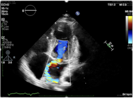
Fifteen years later, the patient developed arterial hypertension, which was successfully managed pharmacologically. Cardiac catheterization revealed a 7.5-mm diameter of the aortic arch distal to the brachiocephalic trunk, resulting in a systolic pressure gradient of 37 mmHg. Four years later, transthoracic echocardiography demonstrated severe mitral regurgitation (Figure 1) and stenosis (Figure 2,3) caused by a typical dysplastic parachute mitral valve with a single papillary muscle (Figure 4), along with signs of severe recurrent coarctation, evidenced by an increased gradient in the ascending aorta (Figure 5).
Repeat cardiac catheterization showed significant narrowing of the aortic arch with a maximal pressure gradient of 42-52 mmHg. To precisely localize the stenosis, computed tomography (CT) angiography was performed, revealing narrowing of the aorta between the brachiocephalic trunk and the left common carotid artery (Figure 6).
Due to the progressive increase in the ascending aortic gradient, severe PMV dysfunction, dysplastic, immobile leaflets and sub valvular apparatus which didn’t qualify for a repair, the patient underwent repeat cardiac surgery. A mechanical mitral valve prosthesis (On-X 23 mm) was implanted, and the hypoplastic segment of the aortic arch was replaced with a 20-mm vascular prosthesis.
Postoperatively, the patient developed complete atrioventricular block, and a permanent pacemaker was implanted on postoperative day 10. One year after surgery, the patient reports feeling well, with improved exercise capacity. Blood pressure values have normalized, and the patient currently does not require antihypertensive therapy.
Shone syndrome is a rare congenital heart disease characterized by a combination of four left-sided obstructive lesions: parachute mitral valve, coarctation of the aorta, subaortic stenosis, and supramitral ring [1]. However, not all patients present with the complete spectrum originally described by Shone. Although long-term survival is generally favorable, management requires a staged and individualized approach due to significant anatomical and clinical variability. The treatment of coarctation of the aorta has been debatable. Although it has recently been reported that transcatheter treatment may be a reasonable alternative, surgical treatment could still be a reliable method [2]. Surgical treatment typically prioritizes relief of left ventricular outflow tract obstruction, while mitral valve interventions are often deferred until later in life [3,4].
Close follow-up, particularly during the transition from pediatric to adult care, along with continuous multimodality imaging assessment, is essential to detect late complications and to determine the optimal timing for future interventions, including mitral valve replacement.